
Fig. 4.
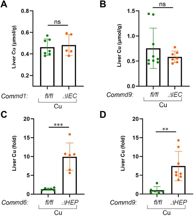
Hepatic but not enteric knockout mice develop altered copper homeostasis. (A) Hepatic copper contents (μmol/g) were measured in dried liver tissue of Commd1ΔIEC mice (n=7) and corresponding floxed control animals (n=5) on a high-copper diet. Results for individual mice are plotted along with the mean and s.e.m. for each group; ns, not significant (unpaired two-tailed Student's t-test). (B) Same analysis as in A, but for Commd9ΔIEC (n=9) and the corresponding littermate controls (n=7). (C) Hepatic copper concentrations were measured in dried liver tissue of Commd6ΔHEP (n=6) and corresponding floxed control animals (n=6) on a high-copper diet. (D) Same analysis as in C, but for Commd9ΔHEP (n=6) and the corresponding littermate controls (n=8). Data are presented as the fold change relative to the average value in the control floxed mouse group. **P<0.01; ***P<0.001 (unpaired two-tailed Student's t-test).