Fig. 2.
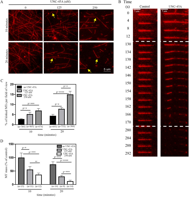
UNC-45A binds, kinks and destabilizes MTs in vitro in a concentration-dependent manner. (A) Sample images of paclitaxel-stabilized MTs in the absence and presence of 125 nM or 250 nM UNC-45A-GFP for 10 min and 20 min. Arrows indicate MTs with kinks. (B) Sample images of time-lapse TIRF microscopy of paclitaxel-stabilized MTs in the presence or absence (control) of 250 nM UNC-45A-GFP. Dashed lines indicate the timeframe in which MTs kink and break. (C) MTs with kinks evaluated in the absence and presence of increasing concentrations of UNC-45A at 10 min and 20 min of incubation. Results are expressed as percentage of kinked MTs per field of view (n=MTs evaluated per condition). (D) MT mass in the absence and presence of increasing concentrations of UNC-45A at 10 min and 20 min of incubation, and determined by measuring the average MT fluorescent intensity from three individual areas per field of view (n=field of view measured per condition). Results are expressed as a percentage of control. *P<0.05, ***P<0.001, ****P<0.0001; ns, not significant.

