Figure 1.

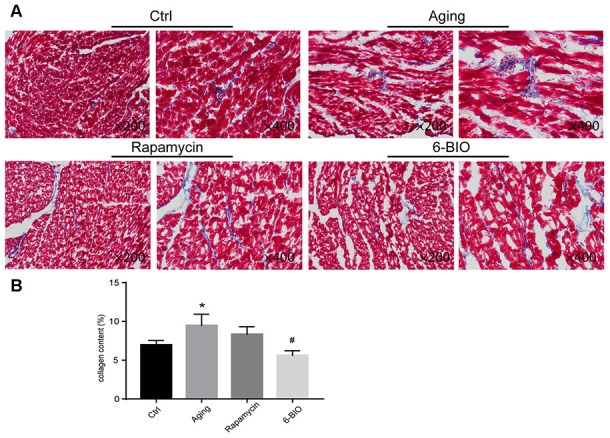
(A, B) Representative photomicrographs of Masson-stained myocardium (×200 and ×400). *P<0.05 compared with the young control group; #P<0.05 compared with the aging group.

(A, B) Representative photomicrographs of Masson-stained myocardium (×200 and ×400). *P<0.05 compared with the young control group; #P<0.05 compared with the aging group.