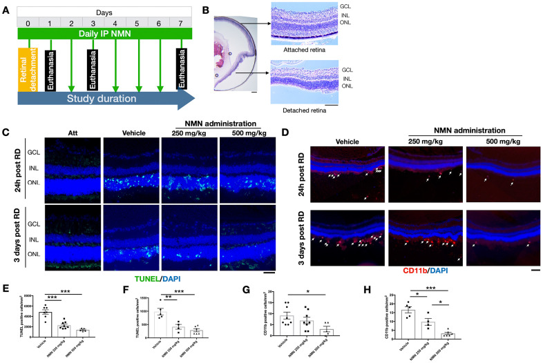
Figure 1.
Protective effects of NMN administration in the early phase of retinal detachment (RD). (A) A flow chart for the in vivo experiments. (B) schematic anatomy of a mouse eye by hematoxylin and eosin (HE) staining showing a bullous after successful RD surgery. Scale bar: 50 μm. (C, E, F) TUNEL+ cells (green) were seen with the highest numbers in vehicle-treated retinas and lowest in the 500 mg/kg NMN-treated retinas at both 24h (E) and three days (F) post RD. Nuclei were counterstained with DAPI (blue). N = 4 to 8 eyes per group. Scale bar: 50 μm. (D, G, H) CD11b+ macrophages (red) infiltrated in the subretinal space after RD, NMN administration significantly reduced the number as soon as 24h post RD (G), and in a dose-dependent manner after three days of RD (H). Nuclei were counterstained with DAPI (blue). N = 5 to 8 eyes per group. Scale bar: 100μm. Statistical significance was analyzed with one-way ANOVA followed by Tukey-Kramer adjustments. *p<0.05, **p<0.01, ***p<0.001. Data are mean ± SEM.

