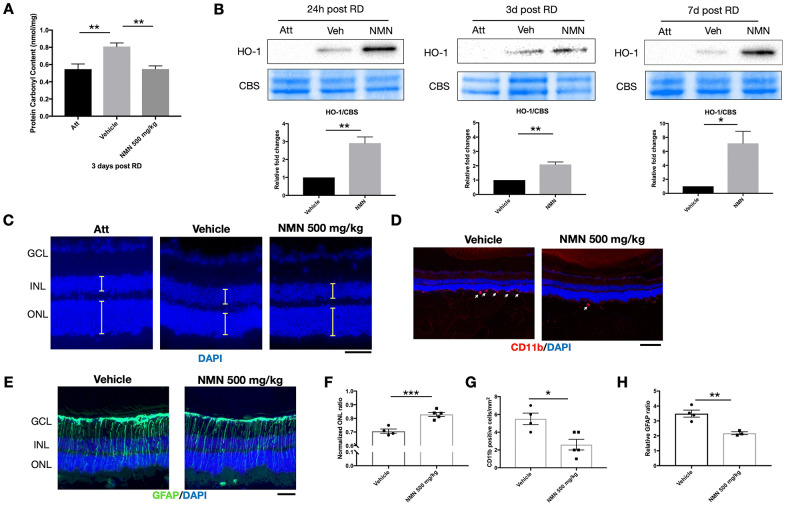
Figure 2.
NMN administration attenuates oxidative stress and is protective in the late phase of retinal detachment (RD). (A) Protein carbonyl content (PCC) was significantly higher after RD while NMN treatment normalized PCC level comparable to the attached retinas at three days post RD. N = 6 eyes per group. (B) Heme oxygenase 1 (HO-1) was significantly up-regulated in the NMN groups as soon as 24h and remained highly expressed throughout the experimental period. N = 4 to 5 eyes per group. (C–F) Preservation of outer nuclear layer (ONL) thickness after NMN administration was seen 7 days after RD. N = 4 to 5 eyes per group. Scale bar=50 μm. The thickness of the layers was measured as the yellow lines indicated. (D–G) Inhibition of neuroinflammation seen by a reduced number of CD11b+ infiltrating macrophages (red) in NMN-treated retinas compared to vehicle-treated retinas. N = 4 to 5 eyes per group. Scale bar: 100 μm. (E–H) Reduced reactive gliosis represented by GFAP (green) staining in NMN-treated retinas compared to vehicle-treated retinas. N = 3 to 4 eyes per group. Scale bar: 50 μm. Statistical significance was analyzed with one-way ANOVA followed by Tukey-Kramer adjustments or the unpaired Student's t-test. *p<0.05. **p<0.01. ***p<0.001. Data are mean ± SEM.

