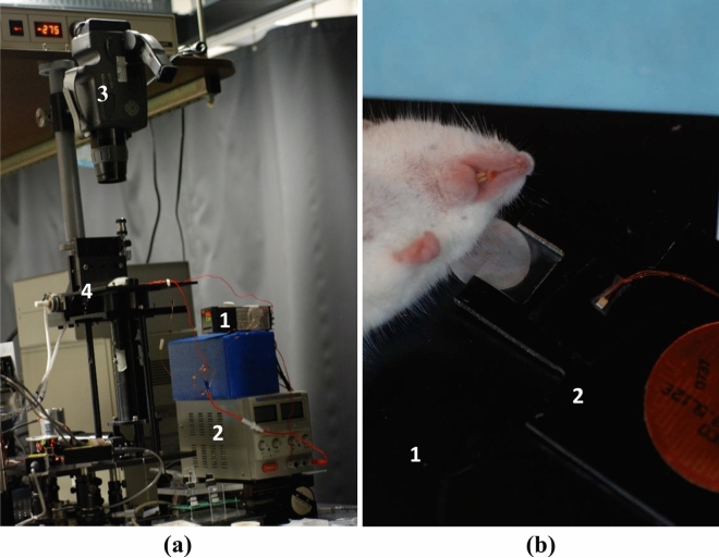
Figure 5.
Imaging experiments lay-out. (a) Reflectance confocal microscope with heating apparatus and thermal camera. External temperature controller (1) and power supply (2) were used to heat and maintain the desired tissue temperature. Thermal camera (3) was positioned above the sample stage (4) to provide independent temperature monitoring. (b) Picture of mouse ear positioning on the sample stage (1) of the confocal microscopy system. Ear tissue was heated using the same heating apparatus (2) as for the integrating sphere measurements.

