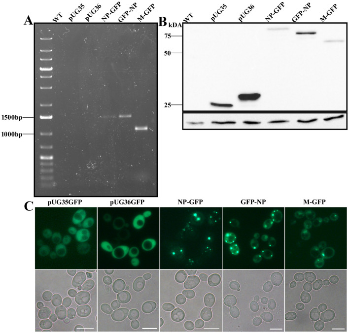
Fig. 1.
Expression of GFP-tagged NDV NP and M proteins in yeast. Yeast RNA was isolated and used for RT-PCR to confirm transcription of viral proteins (a). TCA extracts of cell lysates expressing NDV NP-GFP, GFP-NP, M-GFP, pUG35, pUG36 and WT cells without any plasmid were analyzed by western blotting using α-GFP (b). A single band corresponding to the size of the respective proteins is observed. The panel below represents actin used as a loading control. (c) Fluorescence microscopy images of the cells expressing the above-mentioned plasmids. Upper panel represents GFP signal, and lower panel is the bright field image. Scale bar represents 5 µm

