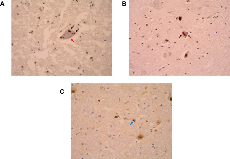
Figure 3.
Neuropathological hallmarks of TDP-43 proteinopathies as observed with TDP-43 immunohistochemistry. (A) An alpha motor neuron in the cervical spinal cord of a patient with amyotrophic lateral sclerosis (ALS) demonstrating intraneuronal cytoplasmic inclusions of TDP-43 (black arrows) and nuclear clearance (nucleus stained blue, red arrow). (B) Intraneuronal cytoplasmic aggregates (black arrow) and nuclear clearance (nucleus stained blue, red arrow) in a neuron of the inferior olivary nucleus. (C) Glial coiled body-like inclusions (blue arrow) in the caudal pons. All images represent light microscopy micrographs obtained at ×400 magnification (University of Newcastle). The antibody used was the proteintech antibody (Proteintech, rabbit polyclonal; product code 1072-2-AP). All images are derived from patients with sporadic ALS.

