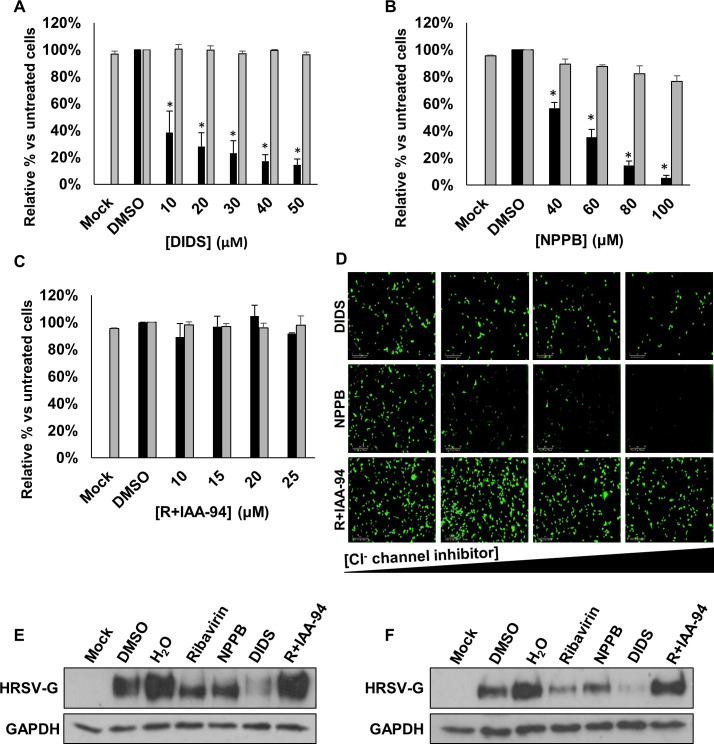
Figure 2.
Broad-spectrum Cl− channel inhibitors DIDS and NPPB decrease HRSV-GFP infection. (A–C) HRSV-GFP expression in A549 cells pretreated with broad-spectrum Cl− channel inhibitors DIDS (10–50 µM), NPPB (40–100 µM) and R+IAA-94 (10–25 µM), shown as a percentage of DMSO control cells (GFP count per well—black bars). Cell viability in each condition was determined via MTS assay (grey bars). Experiments were performed in duplicate in 96-well plates and represent the average of four independent repeats (n=4)±SE. p<0.05 is represented by an asterisk (*). (D) Representative Incucyte images of A549 cells treated with increasing concentrations of DIDS, NPPB and R+IAA-94 (left to right) and infected with HRSV-GFP. Images taken using IncuCyte ZOOM software. Scale bar: 300 µm. (E–F) Western blot analysis of wild type HRSV glycoprotein (G) expression as a marker of infection in A549 (left) or SH-SY5Y cells (right). Cells pretreated with NPPB (60 µM); DIDS (40 µM); R+IAA-94, (40 µM) or the known HRSV inhibitor ribavirin (60 µM) alongside solvent-treated controls (DMSO and H2O). GAPDH was probed as a loading control. DIDS, 4,4ʹ-diisothiocyanostilbene-2,2ʹ-disulfonic acid; NPPB, 5-nitro-2-(3-phenylpropylamino) benzoic acid.

