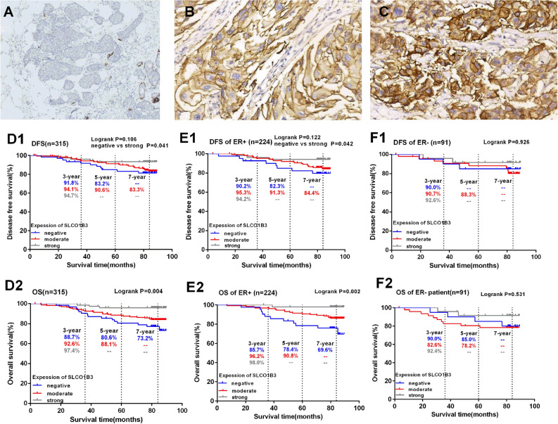
Figure 1.
SLCO1B3 staining in representative tissue specimens, and the association between SLCO1B3 expression and the clinical outcome of patients with primary invasive breast cancer. The immunohistochemistry staining of SLCO1B3 on representative tumor specimens and normal breast tissue are shown. Panel (A) shows the negative SLCO1B3 expression in breast cancer tissue (× 200); panel (B,C) show moderately positive (B) and strongly positive (C) SLCO1B3 expression in the breast cancer tissues (× 200). Kaplan–Meier curves for DFS and OS were plotted based on different SLCO1B3 expression levels in all breast cancer patients (D1,D2), or in ER + breast cancer patients (E1,E2) and ER- breast cancer patients (F1,F2).

