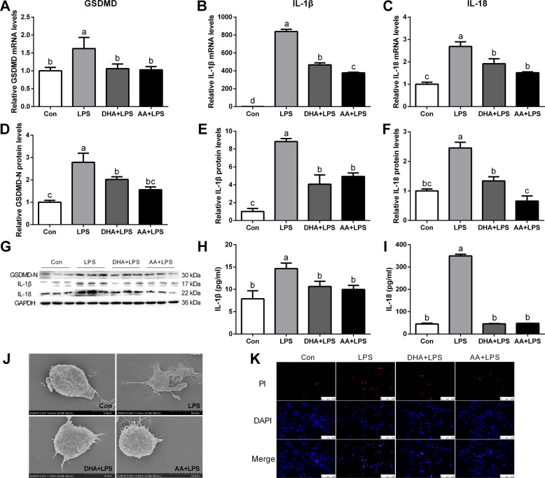
Fig. 2. DHA/AA inhibited the expression of GSDMD, IL-1β and IL-18, and cell inflammatory death in LPS-induced Kupffer cells.
A–C Kupffer cells were pretreated with 50 μM DHA/AA for 4 h and then were treated with 100 ng/mL LPS for 6 h. The mRNA levels of GSDMD, IL-1β, and IL-18 were determined by quantitative real-time PCR. D–G Kupffer cells were pretreated with 50 μM DHA/AA for 4 h and then were treated with 100 ng/mL LPS for 12 h. The protein levels of GSDMD, IL-1β, and IL-18 were determined by western blot analysis. H, I Levels of IL-1β and IL-18 in the supernatant were measured by ELISA. J The cell morphology was observed and imaged under a scanning electron microscope (SEM). K PI staining was used in immunofluorescence (scale bar, 100 μm). In the bar graph, data represent the means ± SEM, n = 3 per group. Mean values not sharing the same letters are significantly different, P < 0.05.

