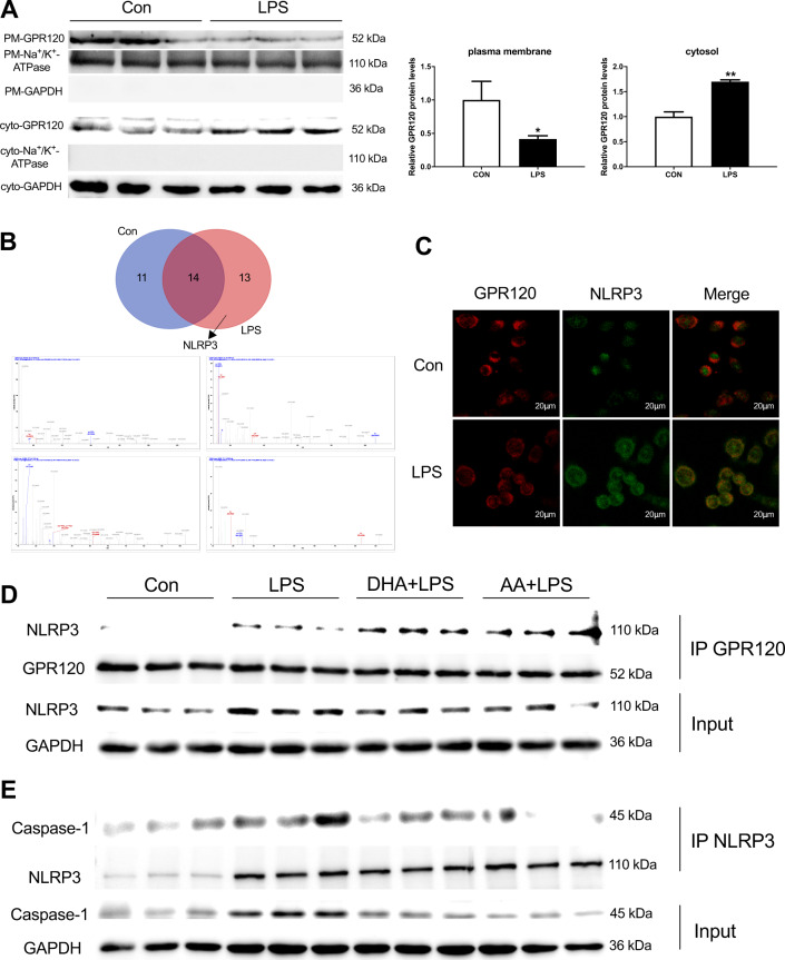
Fig. 4. DHA/AA promoted the interaction between GPR120 and NLRP3 in LPS-induced Kupffer cells.
Kupffer cells were treated with 100 ng/mL LPS for 12 h. A The GPR120 protein expression in the plasma membrane and cytoplasm. B The expression of NLRP3 protein was observed by the Venn diagram and LC-MS/MS analysis (the upper image is the Venn diagram and the lower one is the NLRP3 mass spectrogram). C GPR120 and NLRP3 co-localization was observed in immunofluorescence (scale bar, 20 μm); D, E Kupffer cells were pretreated with 50 μM DHA/AA for 4 h and then treated with 100 ng/mL LPS for 12 h. The cell lysates were immunoprecipitated with GPR120 antibody and NLRP3 antibody separately, and then the samples were analyzed by western blot for GPR120, NLRP3, and caspase-1 as indicated in the text. In the bar graph, data represent the means ± SEM, n = 3 per group. **P < 0.01 and *P < 0.05.

