Figure 4.
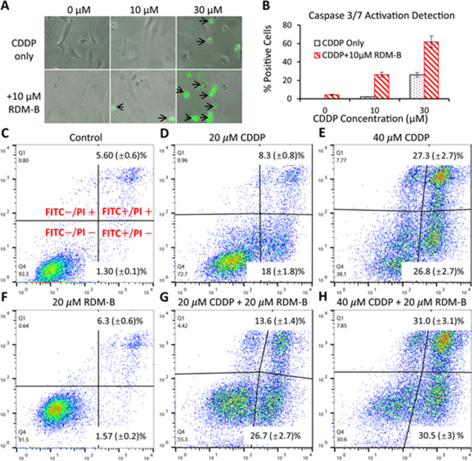
In vitro apoptosis detection in cervical cancer HeLa cells. (A) Representative pictures (black arrows indicating morphological changes) of HeLa cells treated with 10 μM and 30 μM CDDP with/without the combination of 10 μM RDM-B (BV10) for 12 h and then incubated with the CellEvent Caspase-3/7 Green Detection Reagent and analyzed using fluorescence microscopy. (B) Percentages of activated caspases 3/7. (C)–(H) Early/late apoptosis measurements of HeLa cells treated with 20 μM and 40 μM CDDP with/without the combination of 20 μM BV10 for 18 h using an Annexin V-FITC Apoptosis Detection Kit, where the cells were double stained with Annexin-V-FITC and PI. Quantitative analyses of the cell images in A and flow cytometry data in (C)–(H) were performed using an ImageJ software (https://imagej.nih.gov/ij/) and a FlowJo software (https://www.flowjo.com/solutions/flowjo), respectively.

