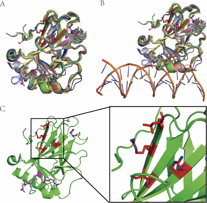
Figure 4.
Identification of allosteric pocket on mutant p53. (A) Three-dimensional (3D) structural alignment of 43 p53 DNA binding domain. (B) DNA binding pocket is composed of fluctuating coli structures (double helix structure represents DNA). (C) Allosteric pocket (red amino acid residues) on p53(4lof) predicted by molecular dynamic simulation and hot spot mutant residues (purple).

