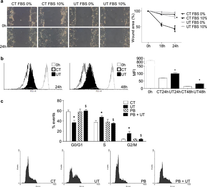
Figure 1.
High doses of UT reduced the proliferation capacity of cultured murine myoblasts, arresting the cell cycle between the phases S and G2/M. C2C12 cells were treated with a mixture of 100 µg/mL IS and 100 µg/mL PC (UT) or vehicle (CT) for 48 h. (a) Scratch wounding was performed on the C2C12 monolayer and wound closure was monitored over the next 24 h in the presence or absence of 10% FBS. Photographs of wounds were captured at 0, 18, and 24 h post-wounding to determine the degree of wound closure. Representative experiment shows only the 0 and 24 h post-wounding photographs. Graph represents the percentage of control (time 0 h) wound area at different times post-wounding from five experiments. *p < 0.05 between CT FBS 10% and UT FBS 10%. (b) Cell proliferation was monitored using CellTrace CFSE solution by flow cytometry during 24 and 48 h. A representative experiment is shown. The bar graph represents the mean fluorescence intensity (MFI). The results are mean ± SEM from five experiments. *p < 0.05 versus CT at the same time. (c) C2C12 cells were treated for 48 h in culture medium with 10% FBS in the presence or absence of 0.25 mM probenecid (PB). Propidium iodide incorporation was assessed by flow cytometry. A representative experiment is shown. The bar graph represents the mean ± SEM from four experiments. *p < 0.05 versus CT. $p < 0.05 versus UT.

