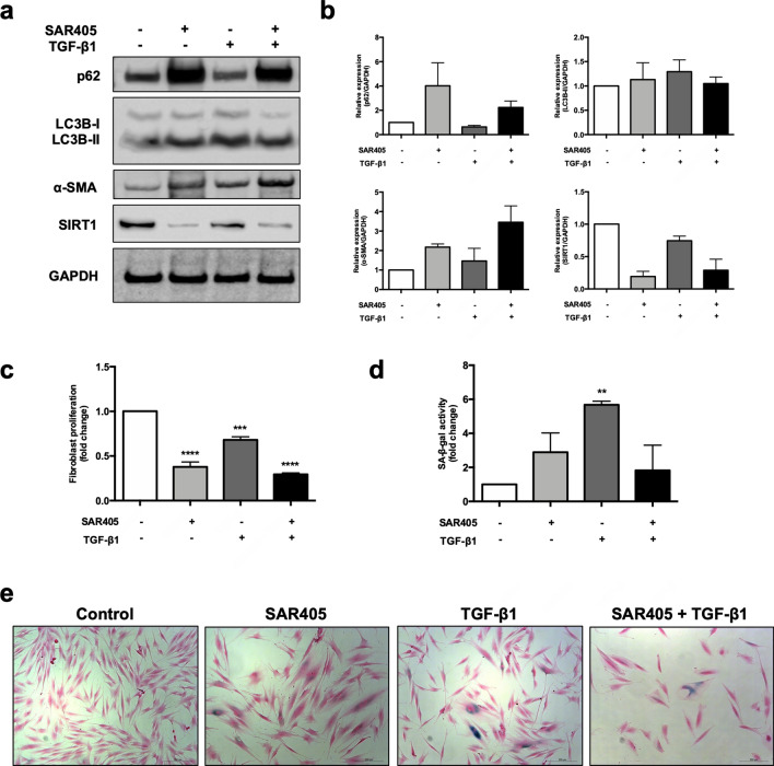
Figure 6.
Autophagic impairment and autophagic flux induce activation and senescence in normal oral fibroblasts. (a) Treatment of NHOF2 with SAR405 for 120 h inhibited autophagy (increased LC3B-II and p62), while TGF-β1 induced autophagic flux (high LC3B-II and low p62). Both SAR405 and TGF-β1 induced the expression of α-SMA and down-regulated SIRT1 levels in NHOF2. Full-length blots are presented in Supplementary Fig. S8. (b) Mean densitometric values from three experiments were normalised to GAPDH and expressed relative to fibroblasts treated with vehicle control (= 1). (c) The proliferation of normal fibroblasts was significantly lower after SAR405 or TGF-β1 treatment as compared to control (= 1). Data are presented as mean ± SEM. ***p < 0.001, ****0.0001, ANOVA compared to control. (d) Fibroblasts with autophagic inhibition (SAR405) and autophagic activation (TGF-β1) displayed a senescent phenotype of higher SA-β-gal positivity. Data are presented as mean ± SEM. **p < 0.01, ANOVA compared to control. (e) Representative images of SA-β-gal staining of SAR405- or/and TGF-β1-treated NHOF2. Treated fibroblasts displayed typical features of senescent cells with enlarged cell size, flat-shaped and contractile myofibroblast-like phenotypes. Scale bar indicates 200 μm.

