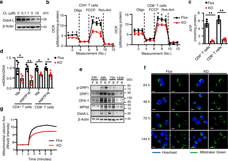
Fig. 2. DsbA-L is a critical regulator of T cell mitochondrial function.
a DsbA-L expression in CD3+ T cells stimulated with different concentrations of CL316,243 for 24 h. b OCR of CD4+ (n = 5/group) and CD8+ (n = 4–5/group) T cells were measured under basal conditions and in response to indicated drugs. Oligo, oligomycin; Rot, rotenone; Ant, antimycin. c ATP production of activated CD4+ (n = 3/group) and CD8+ (n = 3/group) T cells isolated from the spleen of DsbA-LCD4-KO mice and control littermates. d The relative mtDNA content in activated CD4+ (n = 4/group) and CD8+ (n = 4/group) T cells isolated from the spleen of DsbA-LCD4-KO mice and control littermates. e Mitochondrial fission and fusion in activated CD4+ T cells isolated from the spleen of DsbA-LCD4-KO mice and control littermates were measured by western blot analyses. f Mitochondrial morphology was evaluated at indicated time points using live-cell confocal microscopy after staining with 100 nM Mito-Tracker Green and 10 μg/ml Hoechst 33342. Scale bars: 5 μm. g Mitochondrial calcium flux was measured in activated CD4+ T cells isolated from the spleen of DsbA-LCD4-KO mice and control littermates. Results are representative of three independent experiments. Data shown are representative of three independent experiments. All data are presented as mean ± SEM. Statistical values p < 0.05 (*), p < 0.01 (**), p < 0.001 (***) are determined by two-tailed unpaired Student’s t-test. Source data are provided as a Source Data File.

