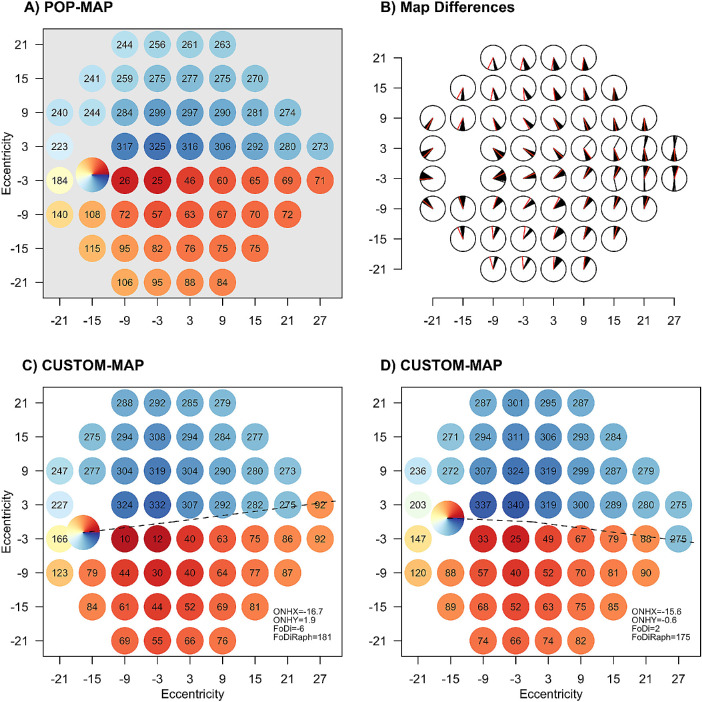
Figure 3.
(A) The population map used in this study (the map of Jansonius et al.,9 with correction of macular locations for Henle fiber length). Within each visual field location, the number responds to the angle of relevance on the optic nerve head with the convention that 0° is temporal, 90° is superior, 180° is nasal, and 270° is inferior. (C and D) Examples of individualized maps for two different eyes with the various anatomic landmarks of relevance from Figure 1 illustrated. (B) A black line for each eye in this study and a red line for POP-MAP show the angle where they map to the optic nerve head for each location in the VF.

