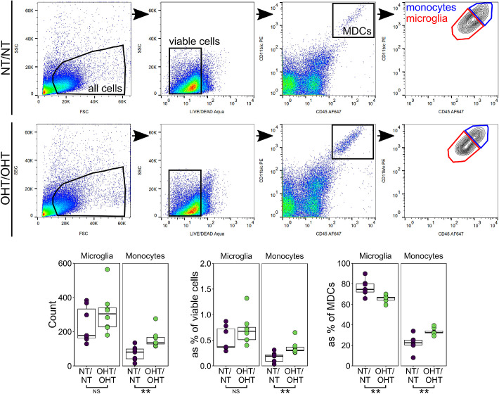
Figure 6.
Monocytes enter the retina early after IOP elevation. To begin to assess whether this increase in microglia number was due to infiltrating monocytes entering the tissue and become microglia-like monocyte derived macrophages, whole retina homogenate was assessed by flow cytometry (n = 7 NT/NT eyes, 8 OHT/OHT eyes). Retinas were sampled at 3 days after injection (the point of peak IOP in this model). At this time point, the microglia numbers were not increased, although there was a significant increase in monocyte numbers (both by raw count and as a percentage of other cell types). MDCs, myeloid derived cells. For complete n please see Methods. *P < 0.05, **P < 0.01, ***P < 0.001. NS, not significant (P > 0.05).

