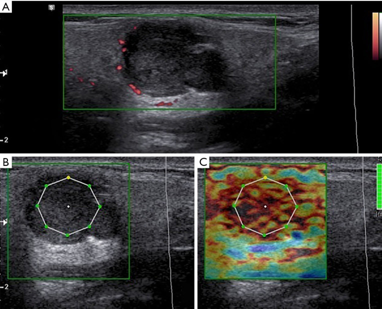
Figure 1.
Grey scale, color Doppler (A) and elastography (B,C) aspect in a patient with pleomorphic adenoma. (A) Iso/hypoechoic lesion of the parotid gland with polycyclic margins, acoustic posterior enhancement and poor peripheral vascular spots but no central flow at color-Doppler US. (B,C) Semiquantitative strain elastography evaluation showed a medium/high stiffness. At the Shear wave elastography the colorimetric pattern of the lesion appears “hard”. The lesion presented high value of ECI at elastography evaluation. It was a pleomorphic adenoma. ECI, elasticity contrast index.

