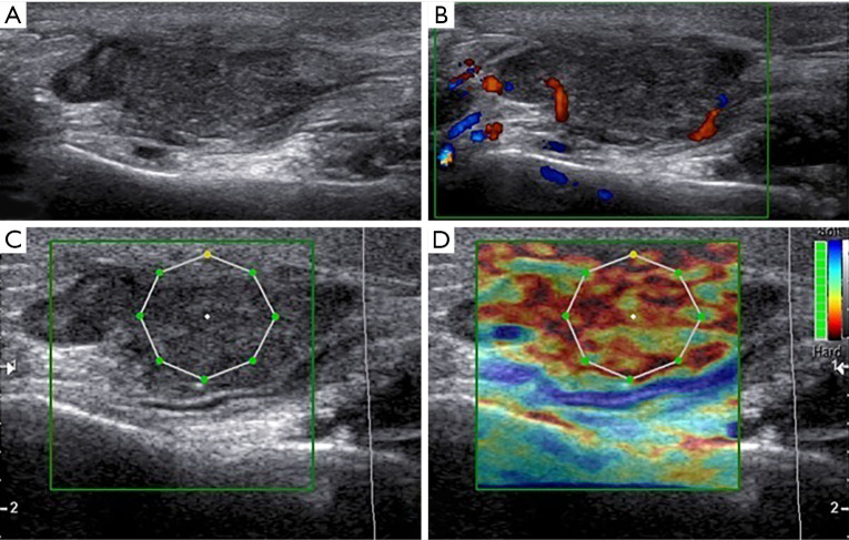
Figure 3.
Grey scale (A), color Doppler (B) and elastography (C) aspect in a patient with a mucoepidermoid carcinoma of the parotid gland. (A) High-grade aggressive lesions showing an irregular structure with hypoechoic inhomogeneous internal architecture, polycyclic margins and posterior acoustic enhancement; (B) at color-Doppler US the lesion showed internal vascular signs and some spots in the peripheral region; (C) the lesion showed high value of ECI at elastography evaluation. The nodule is red on the color map, indicating a “hard” lesion. It was a mucoepidermoid carcinoma of the parotid gland. ECI, elasticity contrast index.

