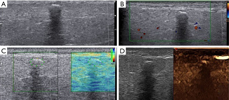
Figure 5.
Grey scale (A), color Doppler (B), elastography (C) and CEUS (D) aspect in a patient with sialoadenitis. (A) B-mode US showed a hypoechoic lesion of parotid gland with small calcific formation that causes posterior wall shadowing. The glandular volume appears increased. The presence of one or more calculi is revealed as bright curvilinear echo complex with acoustic shadowing; (B) color-Doppler didn’t show vascular signs in the lesion; (C) elastosonography presented a low-intermediate ECI value within the lesion. parotid gland with a calculus demonstrated a reduced tissue elasticity; (D) CEUS didn’t show significant enhancement of lesion. It was sialoadenitis. CEUS, contrast-enhanced ultrasound; ECI, elasticity contrast index.

