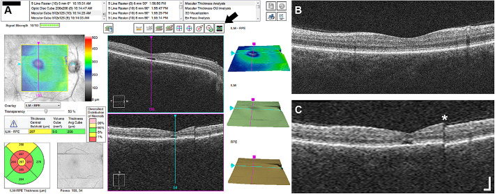
Figure 1.
OCT artifacts in interpolated vertical scans on CIRRUS. (A) Screenshot of the “Macular Thickness Analysis” interface on a CIRRUS HD-OCT device for subject JC_11062. Horizontal B-scans from the macular cube (512 A-scans, 128 B-scans) are displayed in the top middle panel. Interpolated vertical scans from the vertical volume are displayed in the bottom middle panel. The vertical volume is interpolated data between the 128 B-scans in the macular cube volume. Pressing the button noted by the arrow will display the vertical high-resolution B-scan in the bottom middle panel instead of the interpolated vertical scan. (B) Vertical high-resolution B-scan from JC_11062. (C) Interpolated vertical scan (“slow scan”) showing a clear artifact (asterisk) that was not captured in the fast high-resolution scan in B. This vertical volume was assessed as OCT severity category 2. Scale bar: 200 µm.

