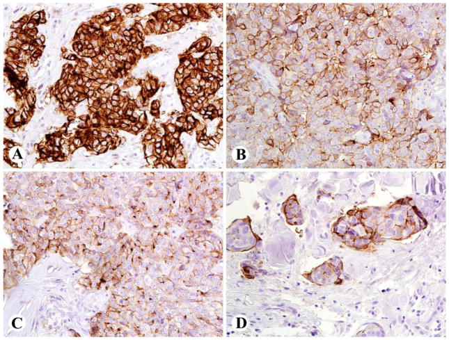
Figure 1.
Patterns of mesothelin immunostaining in thymic non-keratinizing squamous cell carcinomas (SCC) [using monoclonal antibody 5B2 (Novocastra/Leica, Bannockburn, IL) at a dilution of 1:40]. A: Thymic SCC with strong 3+ staining in 100% of tumor cells, with membrane and cytoplasmic labeling in 100% of tumor cells, B: Thymic SCC with strong 3+ staining essentially restricted to cell membranes, in 80% of cells, C: Thymic SCC with moderate to strong 2–3+ staining in cell membrane and cytoplasmic dots, in 70% of cells, D: Thymic SCC with strong membrane staining in 50% of tumor cells.

