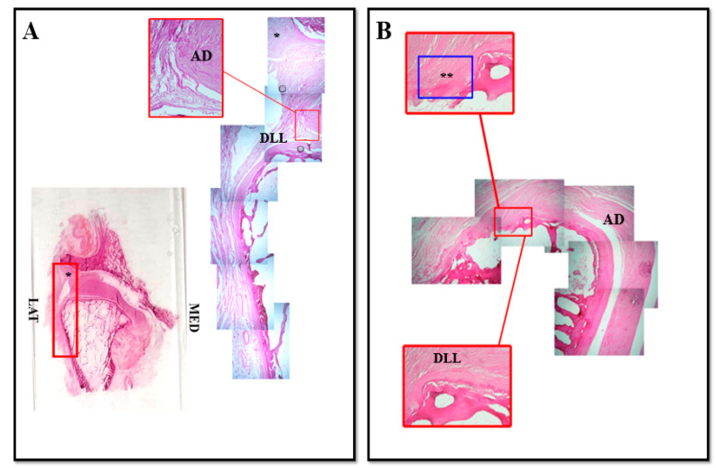
Figure 1.
Compound panel of Hematoxylin-Eosin staining of human TMJ, lateral side (red box). Sections were reconstructed in order to observe a wide microscopic field at the level of the lateral side of TMJ. It is possible to observe the existence of an anatomical structure that made up of nondissociable fibers that seems to give life to two connective bundles: the first one originates from the lateral region of the disc and, running upwards, it inserts on the capsule ((A), asterisk); the second one originates from the lateral region of the disc and it runs downwards inserting on the superficial layer of the articular cartilage of the condyle ((A), DLL). In our opinion this second structure corresponds to the discal lateral ligament; in (B) pictures show the direct insertion of discal lateral ligament on the superficial layer of mandibular condyle cartilage surface (double asterisk). AD: articular disc; DLL: discal lateral ligament; LAT: lateral; MED: medial.

