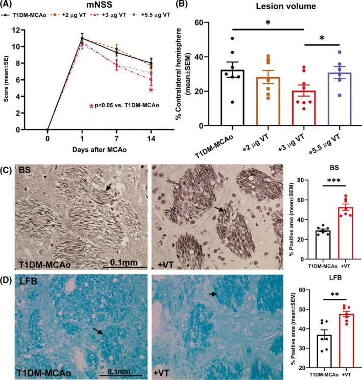
Figure 1.

(A) Neurological function was evaluated on days 1, 7, and 14 after stroke using modified neurological severity score (mNSS) test and analyzed using 2‐way ANOVA and Tukey's post hoc test. Treatment of T1DM stroke with 3 µg/kg VT initiated 24 h after stroke significantly improves neurological functional outcome compared to PBS‐treated T1DM‐MCAo rats. (B) At 14 days after stroke, lesion volume was evaluated using H&E staining and analyzed using unpaired Student's t‐test with Welch's correction. Treatment of T1DM stroke with 3 µg/kg VT initiated 24 h after stroke significantly decreases lesion volume compared to PBS‐treated or 5.5 µg/kg VT‐treated T1DM‐MCAo rats. Therefore, 3 µg/kg VT is identified as a therapeutic dose and used for subsequent analysis. (C‐D) Bielschowsky silver (BS) and Luxol fast blue (LFB) staining were employed to evaluate axon and myelin density and analyzed using unpaired Student's t‐test with Welch's correction. Treatment of T1DM stroke with 3 µg/kg VT significantly increases axon density and myelin density in the striatal white matter in ischemic boundary zone compared to PBS‐treated T1DM‐MCAo rats. Arrows indicate positive staining. N = 6–8 animals per group. *p < 0.05, **p < 0.01, ***p < 0.001
