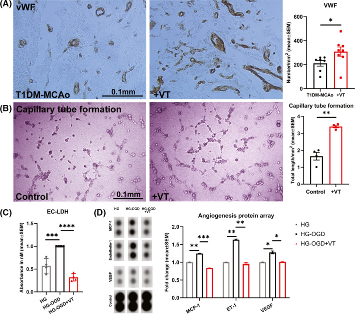
Figure 3.

(A) Immunohistochemical staining for von Willebrand factor (vWF) was used to evaluate vascular density and data analyzed with non parametric, two‐tailed Mann‐Whitney test. Treatment of T1DM stroke with 3 µg/kg VT initiated 24 h after stroke significantly increases vascular density in the ischemic boundary zone compared to PBS‐treated T1DM‐MCAo rats. N = 7–8 animals per group. *p < 0.05. (B) To confirm angiogenic effects of VT treatment in vitro, endothelial cell capillary tube formation assay was employed and analyzed using unpaired Student's t‐test with Welch's correction. Under high glucose (HG) conditions, 10 nM VT treatment significantly increases total length of capillary tube formation, that is, tracks of endothelial cells organized into networks of cellular cords or tubes compared to untreated control group. n = 4 wells/group, **p < 0.01. (C) To evaluate the effect of VT treatment on endothelial cell toxicity, LDH assay followed by one‐way ANOVA and Tukey's post hoc test was performed. Treatment with 10 ng/ml VT significantly decreases endothelial cell toxicity under conditions of HG and oxygen glucose deprivation compared to non‐treated control group. n = 4 wells/group, ***p < 0.001, ****p < 0.0001. (D) To evaluate the differential expression of angiogenic factors by endothelial cells following VT treatment, we employed an angiogenesis protein array and data were analyzed using ImageJ and normalized using internal controls. Under HG conditions, oxygen glucose deprivation significantly increases endothelial cell expression of MCP‐1, Endothelin‐1 (ET‐1) and vascular endothelial growth factor (VEGF) compared to HG alone group. Treatment of endothelial cells subjected to HG and oxygen glucose deprivation with 10 ng/ml VT significantly decreases MCP‐1, ET‐1, and VEGF levels compared to non‐treated control group. *p < 0.05, **p < 0.01, ***p < 0.001
