Figure 5.

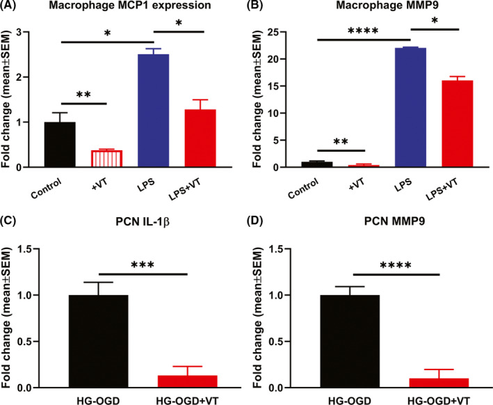
Primary bone marrow derived macrophages were subjected to LPS stimulation to induce M1 polarized macrophage and test whether VT treatment decreases inflammatory factor secretion using RT‐PCR. Macrophages subjected to LPS activation and treated with 10 nM VT exhibit significantly reduced expression of (A) MCP‐1 and (B) MMP9 compared to untreated cells. (C‐D) To test the effect of VT treatment on neuroinflammation, primary cortical neurons (PCNs) were subject to conditions of high glucose (HG) and oxygen glucose deprivation and RT‐PCR was used to evaluate IL‐1β and MMP9 expression. PCNs subjected to conditions of HG and oxygen glucose deprivation and treated with 10 ng/ml VT exhibit significantly decreased expression of inflammatory factors IL‐1β and MMP9 compared to non‐treated control. N = 3/group. *p < 0.05, **p < 0.01, ***p < 0.001, ****p < 0.0001
