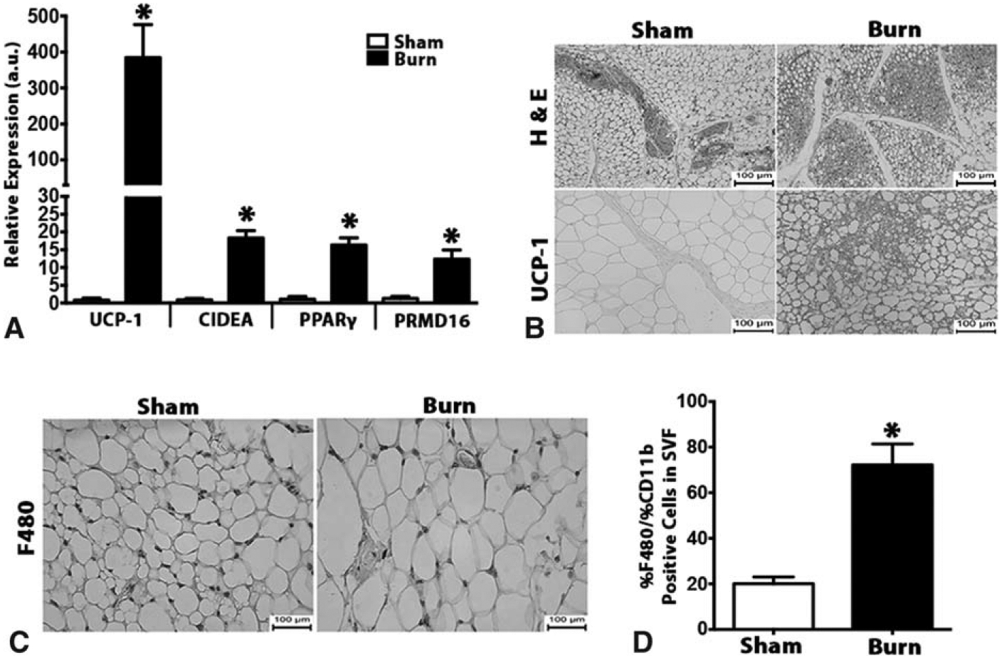
FIGURE 2.

Browning of white adipose tissue in post-burn mice is associated with increased macrophage infiltration. (A) Quantitative RT-PCR analysis of browning genes in inguinal WAT of burned mice and littermate controls. (B) H&E and uncoupling protein 1 (UCP1) staining in inguinal WAT of burned mice and littermate controls. (C) Immunohistochemical staining of macrophage marker F480 in inguinal WAT of burned mice and littermate controls. (D) Flow cytometry analysis of macrophage markers CD11b+F4/80+ cells gated in the inguinal WAT SVF of burned mice and littermate controls. Data are represented as mean ± SEM, P < 0.05. *Significant difference burn vs littermate controls (n = 6).
