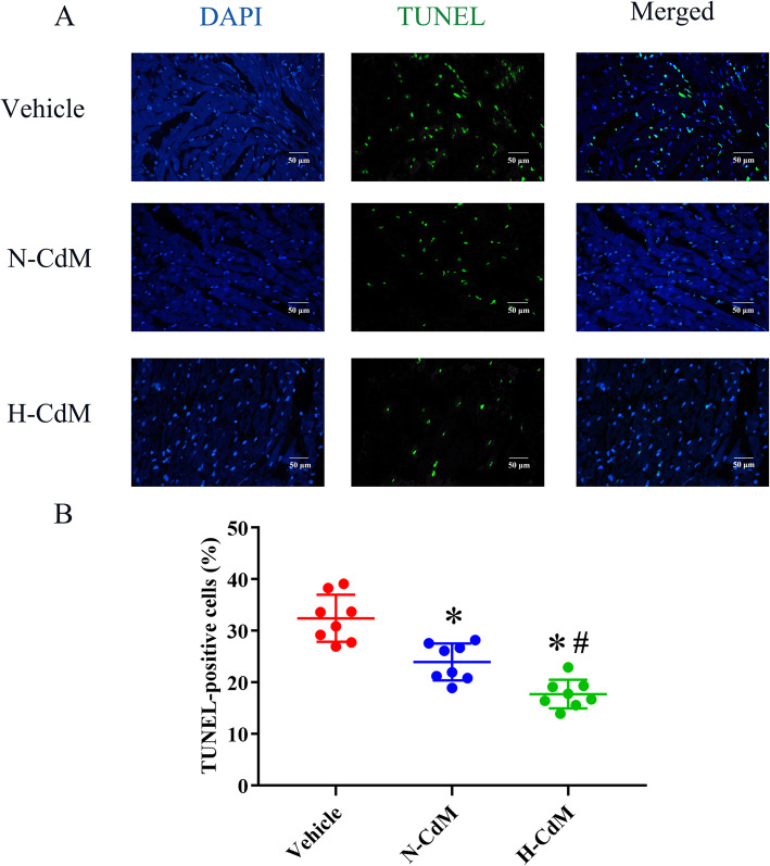
Fig. 6.
Effects of N-CdM and H-CdM on DNA strand breaks in the donor hearts after heart transplantation. a Representative photomicrographs of myocardial tissue stained with 4′,6-diamino-2-phenylindole (DAPI, blue), nuclei with fragmented DNA, as shown by terminal deoxynucleotidyl transferase-mediated dUTP nick end-labeling (TUNEL) staining, and merged image (magnification ×400; scale bar: 50 um). b Quantification of TUNEL-positive cells (as a percentage). N-CdM and H-CdM indicate normoxic and hypoxic conditioned medium, respectively. Data represent mean ± standard error of the mean. One-way ANOVA/Kruskal-Wallis was applied in statistical analysis. *p < 0.05 vs. vehicle, #p < 0.05 vs. N-CdM

