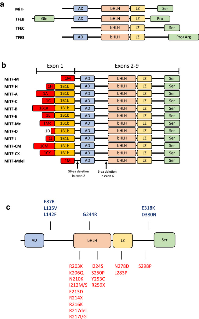
Fig. 1.
a Structure of the four MiT family members. AD, acidic domain; bHLH, basic helix-loop-helix; LZ, leucine zipper; Ser, serine-rich region; Gln, glutamine-rich region; Pro, proline-rich segment; Pro + Arg, proline- and arginine-rich region
(adapted from [1112, 186]. b Different MiTF isoforms. Each isoform is driven by its own promoter and has a partially unique exon, while exons from 2 through 9 are common in all isoforms. c MITF mutations found in melanoma (blue, on top) and in pigment deficiency syndromes (WS2A and TS patients, red, bottom) (adapted from [37]

