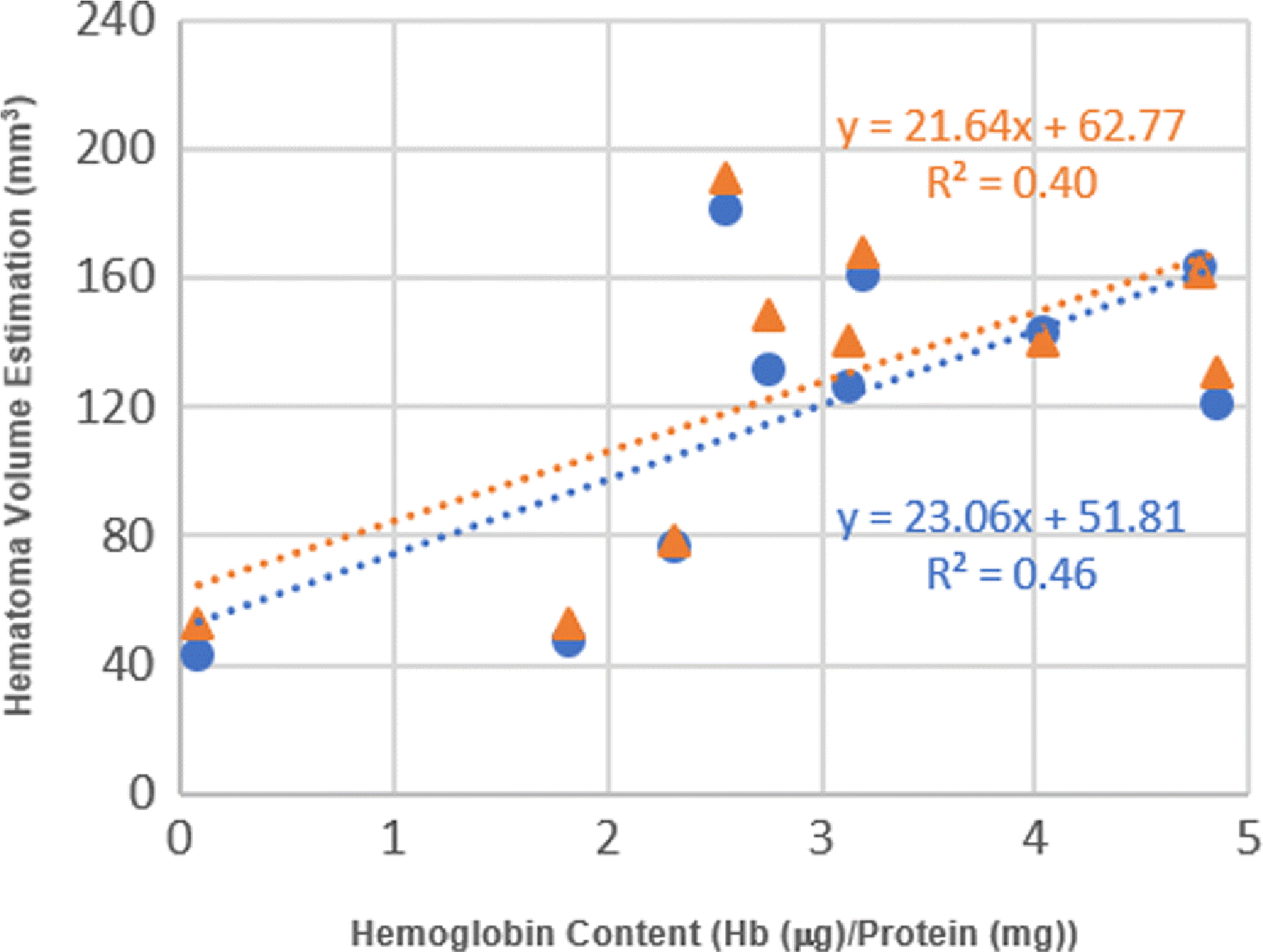
Fig. 5.

Correlation between hemoglobin content and estimated hematoma volume by the manual method and automated method. Data point (orange triangle) shows a linear correlation between hemoglobin content and volume obtained by manual segmentation method (R2 = 0.40, n = 10, p < 0.048). Data point (blue circle) shows a linear correlation between hemoglobin content and volume by automated segmentation method (R2 = 0.46, n = 10, p < 0.031).
