Figure 2.

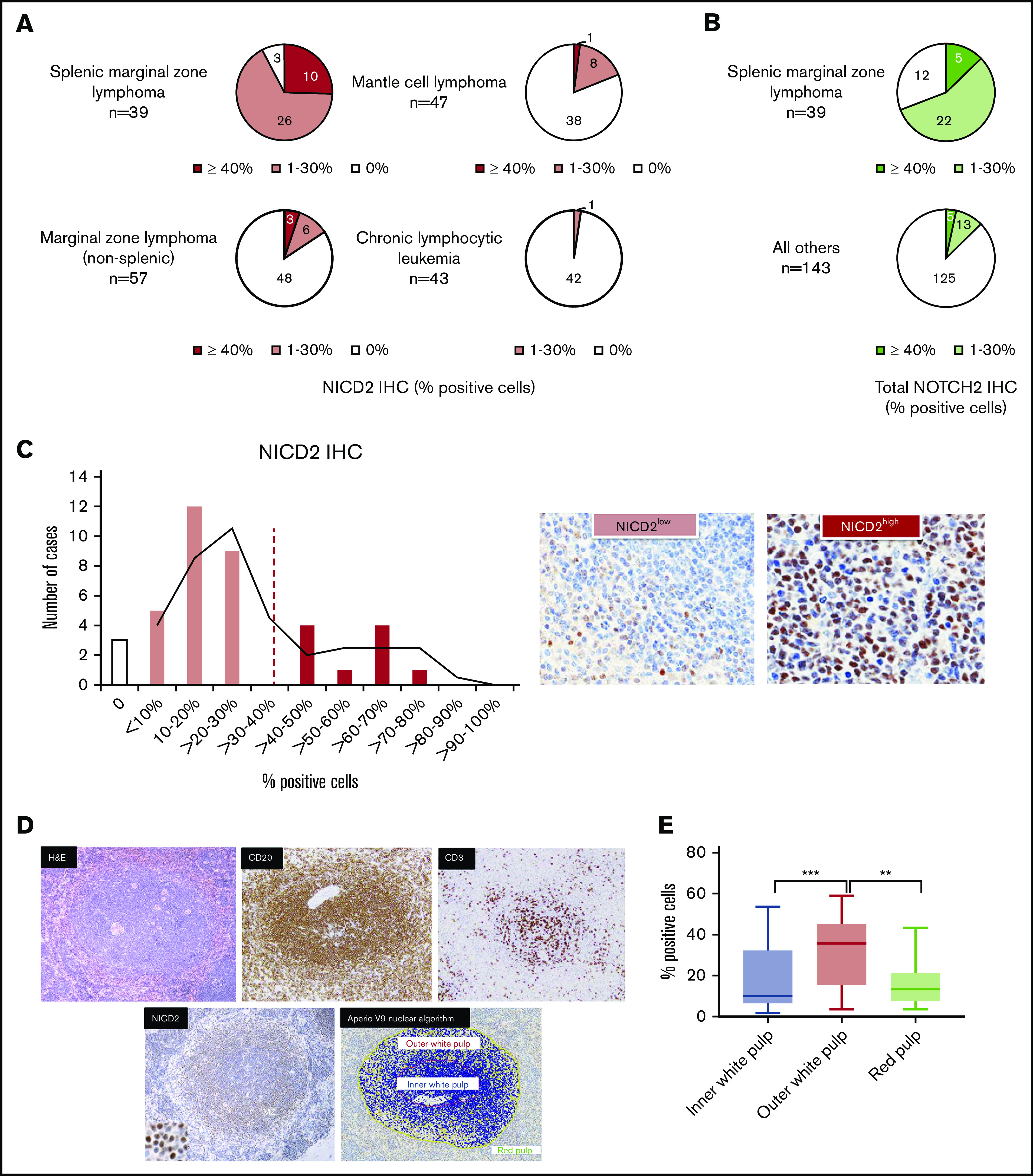
NOTCH2 activation is characteristic of SMZL and is preferentially seen in splenic marginal zones. (A) NICD2 staining in small B-cell lymphomas. (B) Total NOTCH2 staining in small B-cell lymphomas. (C) Histogram showing the percentage of NICD2+ cells across 39 cases of SMZL; representative images of tumors with high (≥40%) and low (<40%) levels of staining are shown. (D) Representative NICD2-stained case of SMZL (hematoxylin and eosin [H&E], CD3, CD20 stains also shown) and image-analyzed depiction of NICD2+ cells in the outer white pulp relative to the inner white pulp (original magnification: ×200; inset, ×400). Scoring was only performed in B-cell–rich areas (defined as >80% B cells). Yellow, positive cells; orange, strongly positive cells; blue, negative cells. (E) Box-and-whisker plots showing the frequency of NICD2+ cells in outer white pulp, red pulp, and inner white pulp in SMZL (***P < .001; **P < .01).
