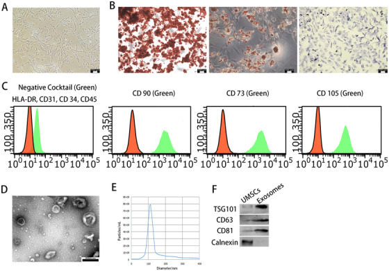
FIGURE 2.

UMSCs were successfully identified and exosomes were successfully extracted. A, Morphological observation of UMSCs (×100). B, UMSCs exhibited multiple osteogenic, adipogenic, and chondrogenic differentiation ability. C, The surface antigen expression of UMSCs was detected by flow cytometric analysis. D, Morphology of exosomes under TEM; scale bar: 200 nm. E, The diameter and concentration of exosomes by nanosight. F, The expressions of TSG101, CD63, CD81, and calnexin in exosomes were detected by Western blot analysis. The data are shown as mean ± SD (n = 3)
