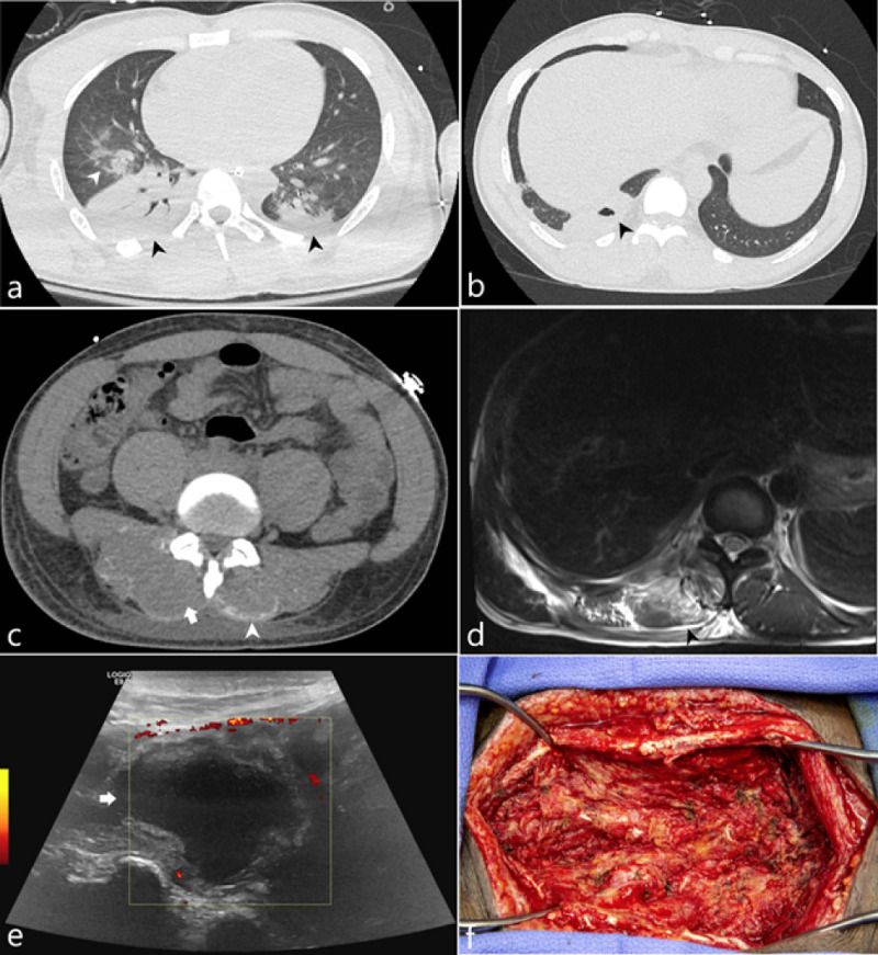
Figure 1. 33-year-old soldier with sickle cell trait was admitted due to rhabdomyolysis complicated by COVID-19 positivity.
(a) Axial CT image of the lung bases acquired on hospital day 3 demonstrates consolidation (black arrowheads) and ground glass opacities (white arrowhead). (b) Axial CT image of the lung base acquired on hospital day 16 demonstrates a cavitation (black arrowhead). (c) Axial CT image of the lumbar spine at the level of the L1 vertebral body on hospital day 16 demonstrates bilateral curvilinear regions of hyperattenuation along the posterior margin of the paraspinal muscles (white arrowhead) as well as a hypoattenuating appearance of the paraspinal muscles. (d) MRI of the lower thoracic spine at the T11 level demonstrates T2 hyperintense signal (black arrowhead) of the paraspinal muscles representing edema. (e) Sonographic evaluation of the paraspinal muscle approximately 43 days from initial admission demonstrates an anechoic fluid collection (white arrow) with a thickened peripheral rim. (f) Operative image of the paraspinal soft tissues status post evacuation.

