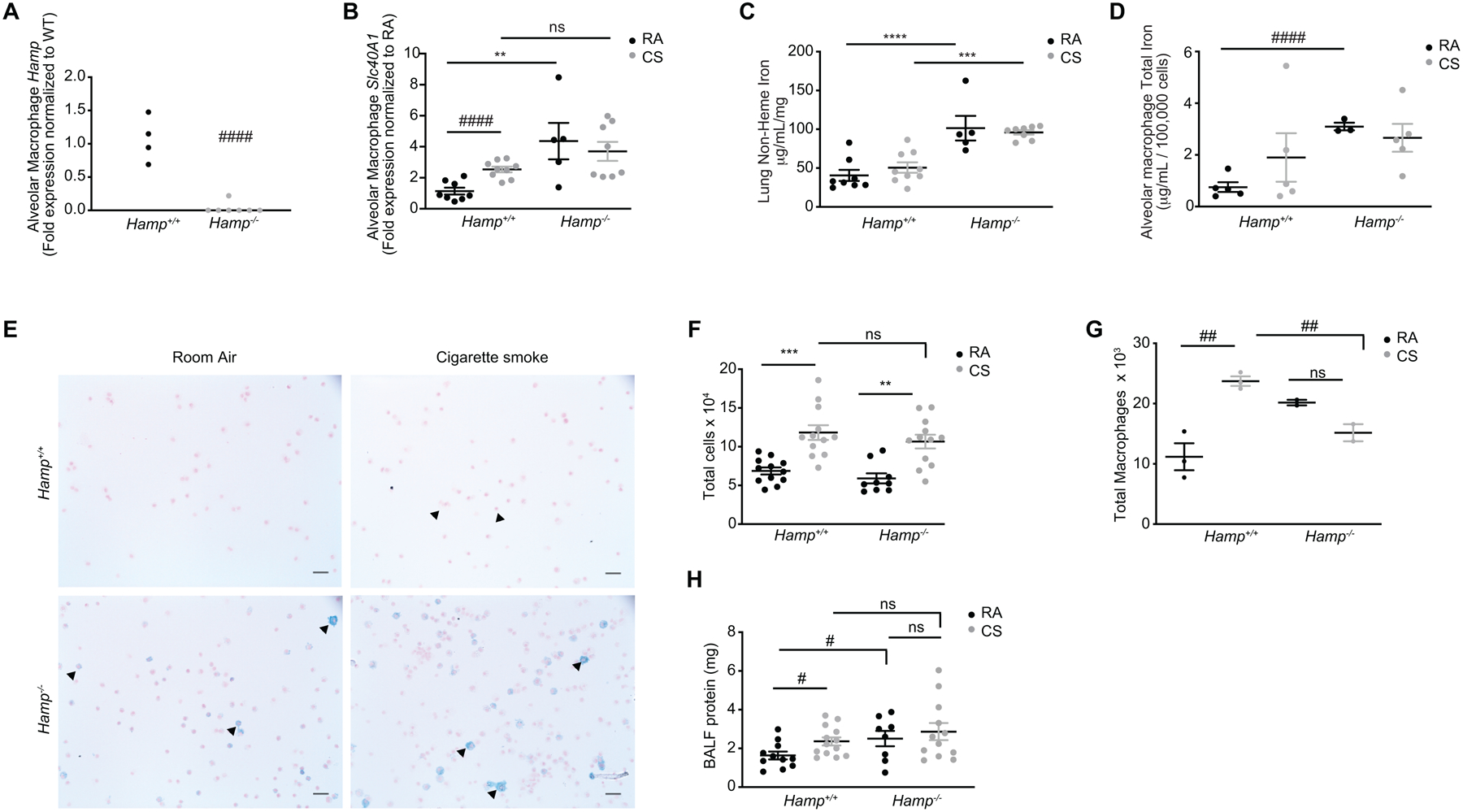
Figure 5. Loss of hepcidin in vivo does not alter smoke-induced injury, iron changes or ferroportin expression.

(a) Hamp mRNA expression in BAL AMs of Hamp+/+ (n = 4) and Hamp−/− (n = 7) mice. (b) Slc40a1 mRNA expression in BAL AMs, (c) non-heme iron levels (μg/ml/mg) in whole lung tissue (excluding BAL cells) of Hamp+/+ (RA n = 8, CS n = 9) and Hamp−/− (RA n = 5, CS n = 8) and (d) total iron levels in BAL AMs of Hamp+/+ (RA, CS n = 5) and Hamp−/− (RA n = 3, CS n = 5) mice exposed to RA or CS (8 weeks). (e) Representative Perls’ staining of BAL AMs of Hamp+/+ and Hamp−/− mice exposed to RA or CS (8 weeks) (arrows indicate presence of ferric iron in alveolar macrophages). (f) Total infiltrating leukocyte cell counts of Hamp+/+ (RA, CS n = 12) and Hamp−/− (RA n = 9, CS n = 12) and (g) total macrophage counts calculated from Hematoxylin-Eosin cytospins of BAL cells of Hamp+/+ (n = 3 per group) and Hamp−/− (n = 2 per group) mice exposed to RA and CS (8 weeks). (h) BALF protein (mg) in Hamp+/+ (RA n = 11, CS n = 13) and Hamp−/− (RA n = 8, CS n = 12) of mice exposed to RA or CS (8 weeks). Scale bars, 50 μm. All data are mean ± s.e.m. **P < 0.01, ***P < 0.005, ****P < 0.001 by one-way ANOVA followed by Tukeys post-hoc test. #P < 0.05, ###P < 0.005 ####P < 0.001 by student’s unpaired t-test. n.s., not significant.
