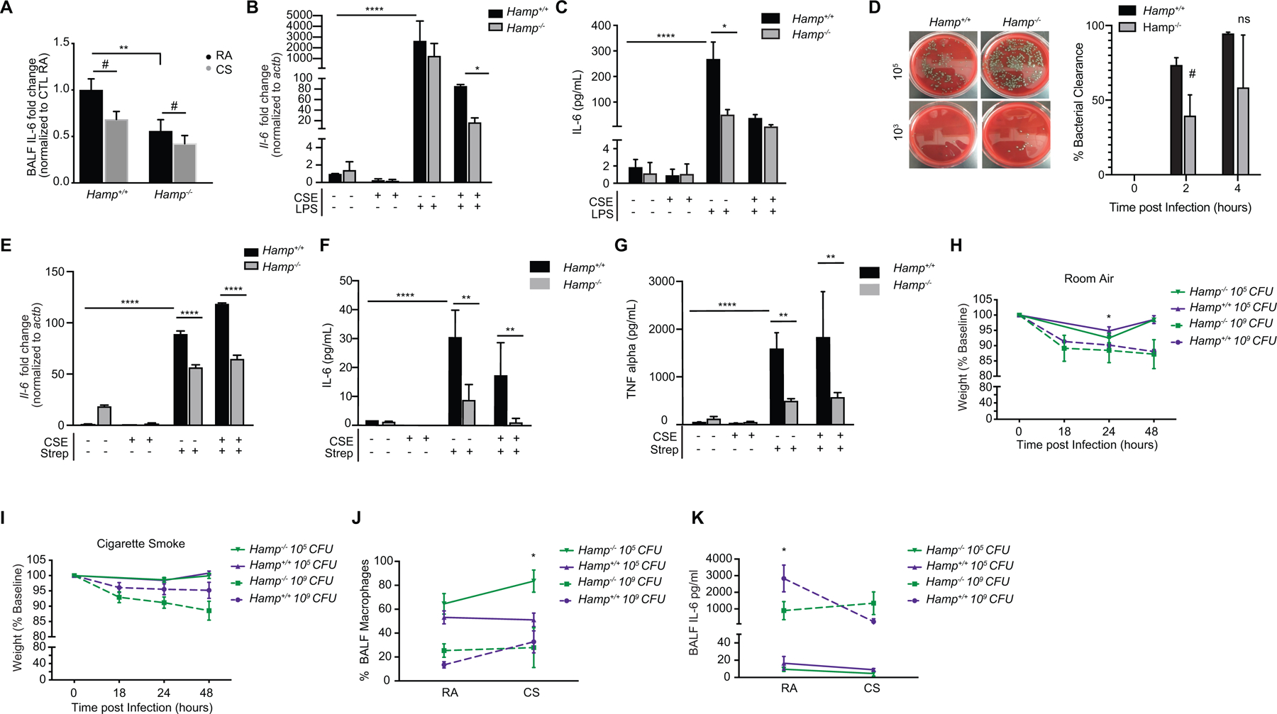
Figure 6. Alveolar macrophages and mice deficient in Hamp have altered immune responses to Streptococcus pneumoniae infection.

(a) Fold change in IL-6 levels in the bronchoalveolar lavage fluid (BALF) of Hamp+/+ (RA n = 8, CS n = 9) and Hamp−/− (RA n = 5, CS n = 8) mice exposed to RA and CS (8 weeks) measured by ELISA. (b) Il-6 mRNA and (c) IL-6 levels (pg/ml by ELISA) in the media of primary AMs isolated from Hamp+/+ and Hamp−/− mice treated with 2% CSE for 18 hours (CSE treatment of 24 hours total) followed by 100 ng/mL LPS (6 hours). Data representative of 3–4 independent experiments. (d) Bacterial titer (left panel) at 103 and 105 dilutions with the number of colony forming units (CFUs) quantified (right panel) in the media supernatants of Hamp+/+and Hamp−/− primary AMs treated with 0.5 ×10⁶ CFU S. pneumoniae for 2 or 4 hours. Data representative of n=4–6 independent experiments. (e) Il-6 mRNA expression (f) IL-6 levels (pg/ml ELISA) and (g) TNF-α levels (pg/ml ELISA) in primary AMs and corresponding media supernatants from Hamp+/+ and Hamp−/− mice treated with 2% CSE (24 hours) followed by S. pneumoniae (4 hours). Data representative of n=2 independent experiments. (h-i) Relative weights, (j) percentage of macrophages in BALF by Hematoxylin-Eosin staining and (k) BALF IL-6 levels (pg/ml ELISA) in Hamp+/+and Hamp−/− (n = 5 per group) mice at 0, 18, 24 and 48 hours post-infection of 105 and 109 (CFU) of S. pneumoniae after exposure to either (h) RA or (i) CS (8 weeks). All data are mean ± s.e.m. *P < 0.05, **P < 0.01, ****P < 0.001 by one-way ANOVA followed by Tukeys post-hoc test. #P < 0.05 by student’s unpaired t-test.
