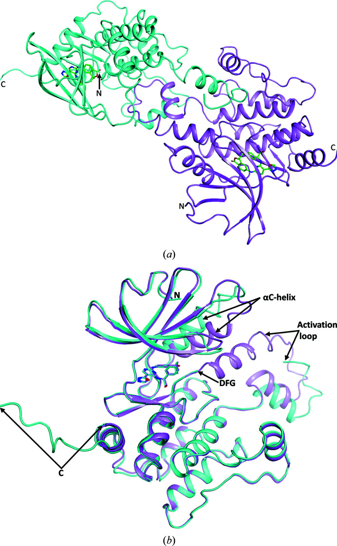
Figure 2.
(a) The contents of the asymmetric unit are shown with chain A in cyan and chain B in violet. Between the two monomers one can see the domain swapping of the activation loop. (b) Superposition of the two monomers (chain A in cyan and chain B in violet) showing that with the exception of the position of the αC-helix and surrounding residues, the activation loop and the extended C-terminus of chain A, the chains follow the same path.

