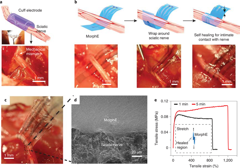
Fig. 2 |. Self-bonding MorphE for soft and conformable neural interfaces.
a, Schematic showing cuff electrode on sciatic nerve. The photographic images showing rigid cuff held by fingers (i) and nerve deformed by the high-modulus cuff electrodes (ii). Scale bar, 1 mm. b, Schematics and images of implantation process for MorphE. Stable enclosure was achieved by wrapping MorphE around the sciatic nerve. Scale bar, 1 mm. c, Image of MorphE being pulled by tweezer after 5 min of self-healing, showing a robust neural interface. Scale bar, 1 mm. d, SEM image (top) showing an intimate device–nerve interface. Scale bar, 20 μm. e, Uniaxial tensile stress–strain curves of two self-healed MorphE at 37 °C in PBS at 5% s−1 strain rate. Before the stress–strain test, a 5 × 5 mm2 overlapped region was attached for 1 and 5 min, respectively to allow self-healing.

