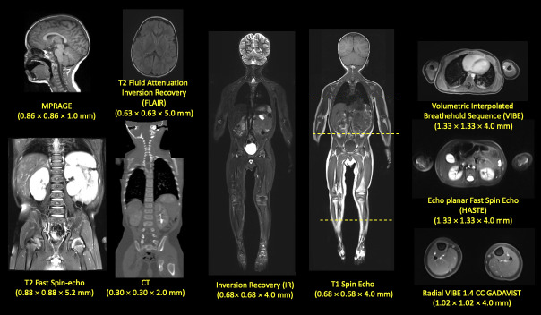
Fig 1. Structural MRI and CT scans used for segmentation.
Several sequences with different resolution and contrast were used for the segmentation of different tissues. For example, MPRAGE, a T1 weighted image, was useful for the segmentation of brain structures while T2 Flair was used for the segmentation of the arteries and veins of the brain and Radial VIBE with a gadolinium-based contrast was used for the segmentation for the vessels of the lower extremities. CT was used for the segmentation of the cortical bone of the pelvis and the core, as well as the base of the skull. Inversion Recovery was useful for the segmentation of the CSF and the Vitreous body of the eyes. HASTE was initially for segmenting the kidneys and the CSF of the spinal cord as well. T2 Fast Spin Echo offered a good outline of the anatomy of the intervertebral disc. All the different tissues were finally referred to the whole body T1 space, while T1 was used for their manual refinement.

