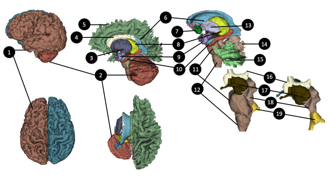
Fig 4. 3D surfaces of the brain and its subcortical areas (created by automatic segmentation algorithm).
1) Left-Cerebral-Cortex, 2) Left-Cerebellum-Cortex, 3) Left-Thalamus, 4) Left-Caudate, 5) Left-Cerebral-White-Matter, 6) Lateral ventricle, 7) Left-Accumbens-area, 8)Left-Putamen, 9)Left-Amygdala, 10) Left-Hippocampus, 11) Pons, 12) Medulla, 13) Left-Pallidum, 14) Vermis, 15) Left-Cerebellum-White-Matter, 16) 3rd-Ventricle, 17) Left-Ventral diencephalon (DC), 18) Midbrain, 19) 4th Ventricle. See S1 Table in S1 File for the list of tissues segmented by an automatic segmentation algorithm [42].

