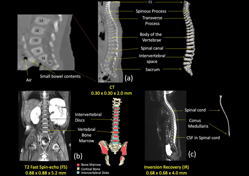
Fig 7. Segmentation of the spinal cord and vertebrae.

(a) Sagittal view of the CT image used for the segmentation of the vertebrae. Arrows on the right were pointing anatomical structures included in the image and represented in the 3D reconstruction while anatomical locations were also marked on the left side. (b) Coronal view of the T2 image used to segment the intervertebral disks and the vertebral bone marrow, as shown with the 3D reconstruction on the right side. (c) Sagittal view of the IR used to delineate the spinal cord and the surrounding cerebrospinal fluid (CSF).
