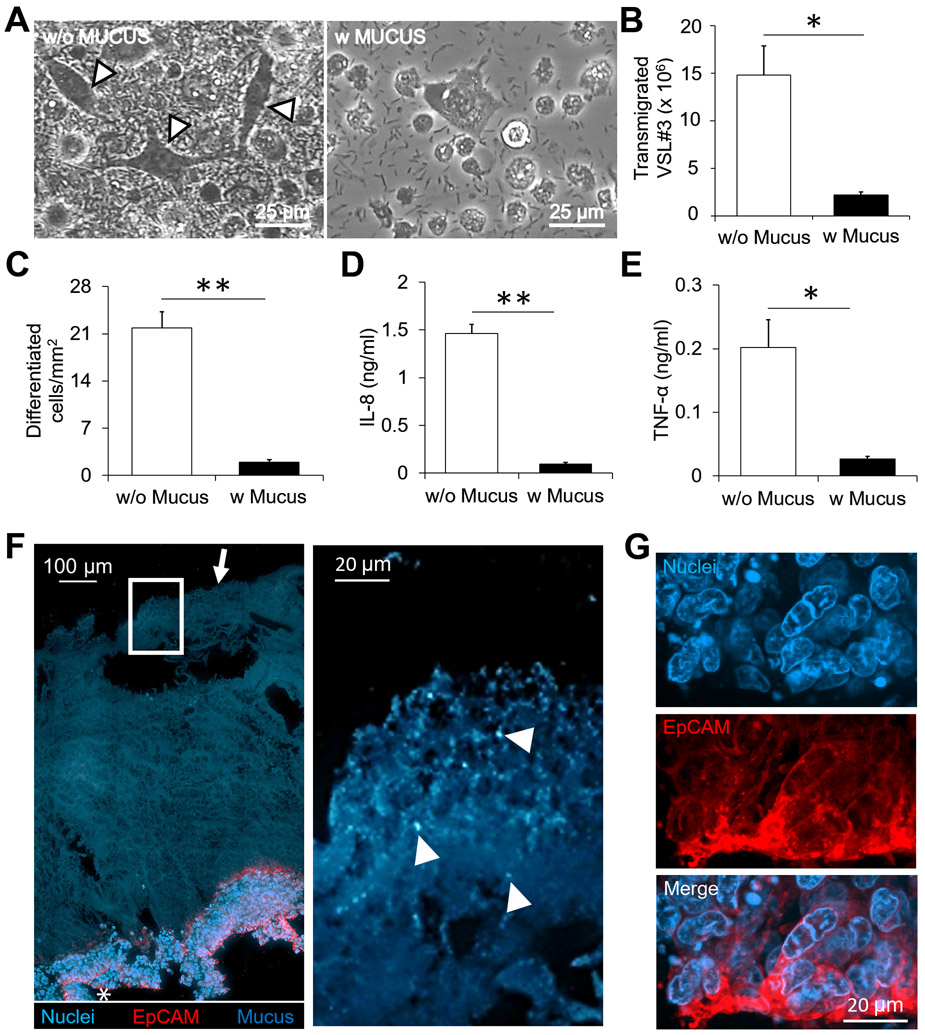
Figure 5. Barrier function and immune modulation by a PSIM gel layer in response to VSL#3.
(A) VSL#3 transmigrated across the epithelial layer and caused THP-1 cell differentiation identified by elongated morphologies (arrows). Images were taken 24 hours after addition of bacteria. (B) The mucus layer significantly reduced the number of transmigrated VSL#3 bacteria (*, P < 0.05; n = 3). (C-E) The mucus layer significantly reduced the number of differentiated cells per mm2 (C; **, P < 0.01; n = 3), the concentration of IL-8 (D; (**, P < 0.01; n = 3), and the concentration of TNF- α (E; *, P < 0.05; n = 3) in the co-culture. (F) A 20 μm thick histological section (Left) stained with EpCAM (Red) and DAPI (Blue). The PSIM gel layer is auto fluorescent (Blue, arrow). Expanded image (Right) shows DAPI-stained VSL#3 bacteria embedded at the outer surface of the PSIM gel layer (Arrows). (G) Expanded images of the epithelial layer (white star) showing nuclei and EpCAM staining.

