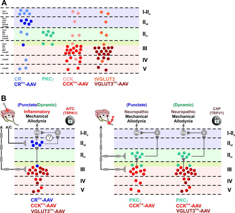
Figure 8. Dorsal horn circuitry underlying mechanical allodynia depends on the injury type.
(A) Schematic of the laminar distribution of virally targeted (AAV) and lineage-derived populations discussed in this study. Virally targeted CR neurons (dark blue) are mostly restricted to the dorsal-inner part of lamina II (IIid) and largely exclude other CR-lineage neurons (light blue) in lamina I, the deep layers, and those expressing PKCγ. Nearly all PKCγ neurons were targeted by the virus (light green). Virally targeted CCK neurons (dark red) are highly concentrated in lamina III with scattered cells in laminae IV-V and generally exclude CCK-lineage neurons in the superficial laminae including those expressing PKCγ (light red). Virally targeted tVGLUT3 neurons (dark brown) are highly concentrated in lamina III with scattered cells in laminae IV-V and exclude tVGLUT3-lineage neurons in superficial laminae including those expressing PKCγ (light brown).
(B) Schematic diagram of the mechanical allodynia circuitry in the context of injury. Targeted CR neurons receive A- and C-fiber input whereas targeted CCK/tVGLUT3 neurons receive predominantly A-fiber input. Left circuit: After inflammation, punctate or dynamic mechanical allodynia engage neurons expressing CCK/tVGLUT3 in lamina III, which activate targeted CR neurons in lamina IIid. These CR neurons ultimately activate projection neurons (PN) directly, or potentially indirectly through vertical cells, to elicit mechanical allodynia. A similar circuit is observed after activating TRPA1 through peripheral injection of mustard oil (AITC). Right circuit: After nerve injury, punctate mechanical allodynia engages CCK, but not tVGLUT3 neurons in lamina III. These neurons then activate PKCγ neurons in lamina IIid and ultimately projection neurons (PN) indirectly through vertical cells. A similar circuit is observed for dynamic mechanical allodynia, but also includes targeted CCK/tVGLUT3 neurons in lamina III. Mechanical allodynia induced by peripheral injection of the TRPV1 agonist capsaicin (CAP) also relies on PKCγ and CCK neurons.

