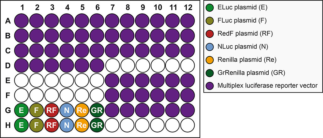
Figure 6. Plate schematic for transfection of mammalian A459 cells.
Wells seeded with cells (see Figure 4) are transfected with plasmid sample: bright green, dull green, red, blue, orange and dark green wells are transfected with constitutively expressing ELuc (E), FLuc (F), RedF (RF), NLuc (NL), Renilla (Re), or GrRenilla (GR) luciferase reporter vectors, respectively, used to determine the transmission coefficients (see Figure 8), while purple wells are transfected with the multiplex luciferase reporter vector containing five pathway transcriptional luciferase reporters (FLuc, RedF, NLuc, Renilla, and GrRenilla) and one control luciferase reporter (ELuc), used to measure pathway-specific manipulations (see Figure 12).

