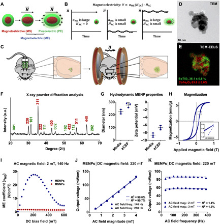
Fig. 1. Material and magnetoelectric characterization of MENPs made from magnetostrictive and piezoelectric phases demonstrates wireless electric field generation.

Schematic demonstrating two-phase magnetoelectricity in materials made from magnetostrictive and piezoelectric materials that are strain-coupled (A). Schematic demonstrating the rationale for using a large DC magnetic field overlaid with an AC field to generate optimal magnetoelectric output (B). Diagram of method of in vivo MENP administration. MENPs are injected bilaterally into the subthalamic region of mice, and MENPs are wirelessly stimulated using an AC and DC magnetic field (C). Transmission electron microscope (TEM) (D) and TEM–electron energy loss spectroscopy (TEM-EELS) images (E) show MENP morphology and BaTiO3/CoFe2O4 phases (green and red, respectively), with quantitative elemental analysis measurement of the molar percentage of each material (E). MENPs were analyzed via x-ray powder diffraction (XRD) to confirm the perovskite crystal structure of BaTiO3 (green) and the spinel crystal structure of CoFe2O4 (red) (F). a.u., arbitrary units. Dynamic light scattering (DLS) was used to characterize MENP hydrodynamic properties in cell culture media and artificial cerebrospinal fluid (aCSF) (G). Magnetization of MENPs was measured over a range of −1 to 1 T, as well as oscillated over a range of 0.205 to 0.235 T (inset) (H). emu, electromagnetic unit. The input-output magnetoelectric coefficient (αME) of particles in a sintered pellet was measured as a function of DC bias field in MENPs and MSNPs (I). Voltage normalized to pellet thickness of MENPs was measured using a 220-mT DC field while varying AC field magnitude (J). The AC field frequency dependence of αME was measured using a 220-mT DC field (K). Plots show individual points with means ± SD (n = 3) (G) and individual points fitted to a linear correlation (J and K).
