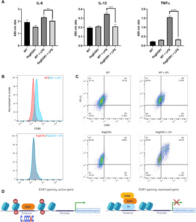
Fig. 6. EGR1 blunts the inflammatory response in human macrophages.

(A) Absorbance values of a cytokine panel ELISA show reduced secretion of select cytokines in EGR1-overexpressing macrophages upon stimulation with LPS. Of the cytokines assayed, IL-6 levels were significantly decreased by ~13%, while IL-12 and TNFα levels were decreased by ~40 and 80%, respectively, as compared to control macrophages after LPS stimulation (**P < 0.01, ***P < 0.001, and ****P < 0.0001). (B) Flow cytometry results of EGR1-overexpressing macrophages exhibit a lack of surface expression of the costimulatory signaling marker CD86 as compared to control macrophages after LPS stimulation. (C) Flow cytometry further shows a distinct population of PDL1(CD274)+- and CD80+-coexpressing cells in EGR1-overexpressing macrophages as compared to control macrophages. (D) Diagrammatic representation of the role of EGR1 in monomacrophagic cells. EGR1 associates with enhancers of both developmental and proinflammatory genes. At developmental enhancers, EGR1 is recruited by its DNA motif and promotes activation, especially during early monocytic commitment (12). Conversely, EGR1 is indirectly recruited at proinflammatory enhancers (likely via repressive TFs) during macrophage differentiation. EGR1 recruitment at inflammatory enhancers leads to deacetylation and loss of accessibility by way of the NuRD-repressive complex.
