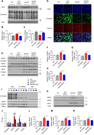
Fig. 2. OGA haploinsufficiency increased global O-GlcNAc levels in the brain and decreased activation of necroptosis.

(A) Western blot analysis of OGA and O-GlcNAcylated proteins in brain samples of indicated mouse genotypes (n = 3). (B and C) Quantification of OGA protein (B) and O-GlcNAc protein (C) level in (A). (D) Immunostaining of O-GlcNAc and P-MLKL in the cortical region of mice samples (n = 3 to 4). DAPI, 4′,6-diamidino-2-phenylindole. Scale bar, 20 μm. (E) Necroptosis-related proteins in the brain of indicated genotypes of mice (n = 3). (F to H) Quantification of P-RIPK1 (F), P-RIPK3 (G), and P-MLKL (H) in (E). The levels of phosphorylated necroptosis–related proteins were normalized to the levels of the corresponding total proteins. (I) Western blot analysis of necroptosis-related proteins in RIPK3 immunoprecipitates from mouse brain samples. The precipitated immunoglobulin G (IgG) heavy chain is marked with an asterisk. IP, immunoprecipitation. (J) Quantification of O-GlcNAc, RIPK1, P-RIPK1, and RIPK3 binding to RIPK3 in (I). (K) Necroptosis-related proteins in insoluble fractions of brain samples collected from the indicated mouse genotypes (n = 3). (L to N) Quantification of necroptosis-related proteins RIPK1 (L), RIPK3 (M), and MLKL (N) in (K). Three slices of each sample were used to normalize each sample. Values are presented as means ± SEM. #P < 0.05, ##P < 0.01, and ###P < 0.001 versus WT; *P < 0.05 and **P < 0.01 versus 5xFAD; one-way analysis of variance (ANOVA) with Tukey’s test.
