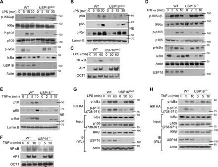
Fig. 3. IKK-induced p105 phosphorylation requires the participation of USP16.

(A) The phosphorylation of IKKs and their substrates in WLs of WT and USP16-deficient BMDMs was measured by IB analysis. (B) The amounts of each NF-κB member in cytoplasmic (CE) and nuclear (NE) extracts were detected by IB. (C) Electrophoretic mobility shift assay (EMSA) of NE extracts of WT and USP16-deficient BMDMs stimulated with LPS (1 μg/ml), as assessed with HRP-labeled NF-κB, AP1, or OCT1. (D to F) Similar analyses of the activation of IKKs (D) and transcription factors (E) were performed via IB and EMSA (E) in USP16−/− MEFs as described above. (G and H) IKK kinase assays and IB assays using WLs of LPS-stimulated BMDMs derived from WT and USP16MKO mice (G) or USP16−/− MEFs (H). The data are representative of at least three independent experiments.
