. 2020 Dec 30;9:e60432. doi: 10.7554/eLife.60432

Figure 7. Nocodazole disrupts Rock and Roll and positional acquisition of hair cells (HCs).

(A) Nascent sibling HCs in myo6b:Yfp-tubulin; emx2:Gfp larvae were treated with nocodazole for 90 min immediately after the HC precursor was observed to divide under live-imaging (0 min). After nocodazole removal, the nascent HC pair started to roll and they exchanged positions (96–174 min). The HC located at the anterior (pink dot) showed stronger Gfp signal due to the presence of emx2:Gfp and Yfp-tubulin alleles (222–282 min, arrowheads) than the emx2:Gfp-negative HC at the posterior (blue dot), which only expresses Yfp-tubulin. (B) Nascent HCs were treated with nocodazole for 150 min after HC precursor divided (0–150 min). After nocodazole removal, HCs failed to exchange their positions (156–312 min), which resulted in the mislocation of the emx2:Gfp-positive HC to the posterior (arrowheads, 618–882 min, pink dots).

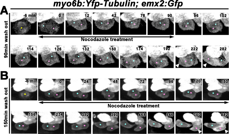
Figure 7.

Figure 7—video 1. Time-lapse video of nocodazole treatment for 90 min of the myo6b:Yfp-Tubulin; emx2:Gfp hair cell (HC) pair shown in Figure 7A.
Download video file (615.4KB, mp4)
Nocodazole was added at 0 min immediately after the HC precursor (−6 min, yellow dot) divided and they were incubated with Nocodazole for 90 min (0–90 min). After removal of nocodazole, the two HCs initiated roll movements to exchange positions with each other (96–174 min). emx2 reporter positive HC (pink dot) was evident as the brighter GFP signal expressing both YFP-tubulin and GFP driven by the emx2 promoter versus the emx2 reporter negative HC (blue dot) expressing only YFP-tubulin (222–282 min).
Figure 7—video 2. Time-lapse video of nocodazole treatment for 150 min of the myo6b:Yfp-Tubulin; emx2:Gfp hair cell (HC) pair shown in Figure 7B.
Download video file (1.4MB, mp4)
Nocodazole was added at 0 min immediately after the HC precursor (−6 min, yellow dot) divided and they were incubated with nocodazole for 150 min (0–150 min). After nocodazole removal, the HC pair did not undergo the Roll phase to exchange their positions. As a result, the brighter GFP-positive, Emx2 HC (pink dot) was mispositioned in the posterior position, whereas the emx2 reporter-negative HC (blue dot) was located in the anterior position (750–882 min).